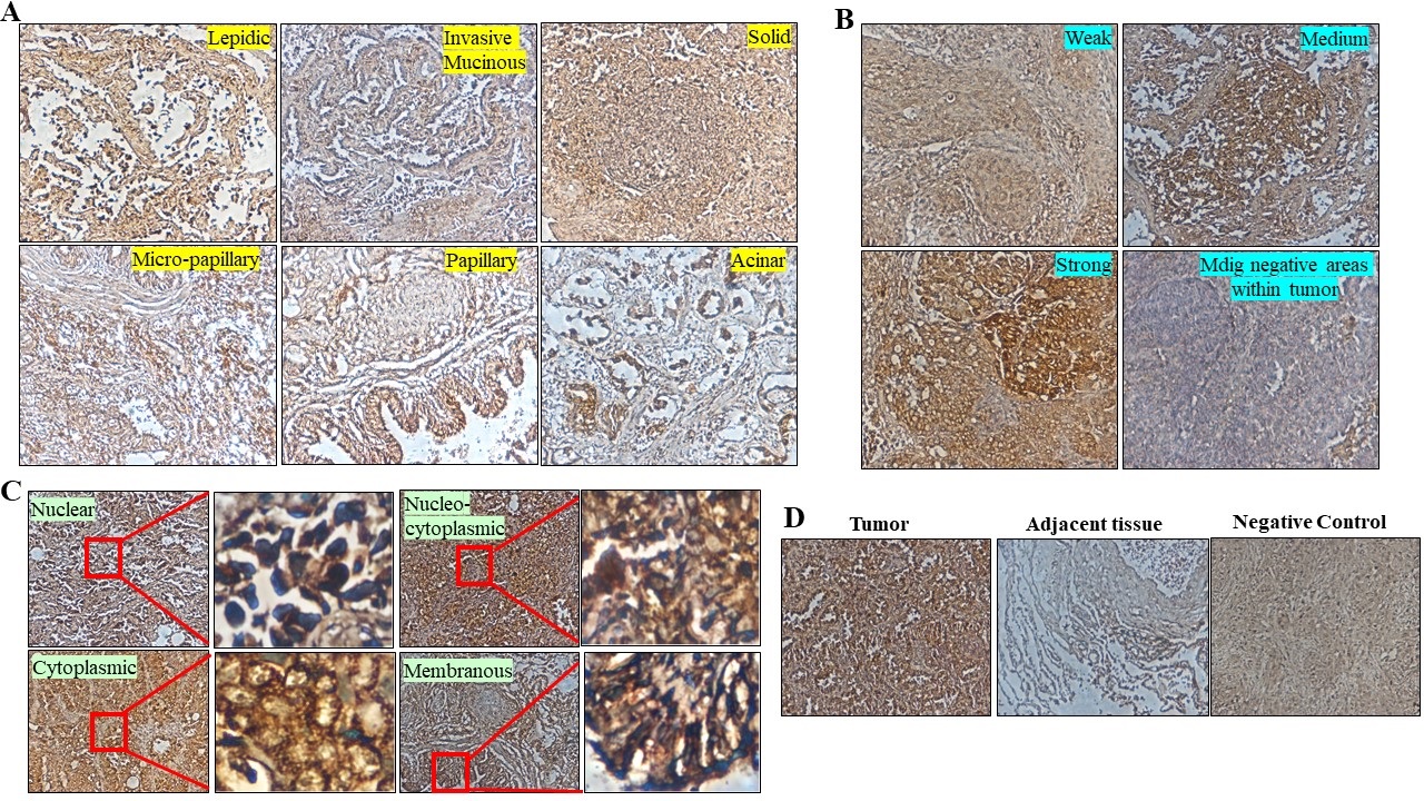
Fig. 4. mdig expression in SMPLC. (A) Six distinct types of histological growth patterns were observed in SMPLC that also stained positive for mdig. Microscopic description of the observed growth patterns in SMPLC tissues reveals a lepidic pattern, where the tumor comprises neoplastic cells lining the alveolar wall with no architectural disruption or stromal, vascular, or pleural invasion. Solid compact nest and sheets of tumor that lack acini, tubules, and papillae constituting the solid growth pattern. In the micropapillary pattern, ill-defined tufts or projections with peripheral nuclei constitute the growth area with no fibrovascular core. However, in the papillary pattern, fibrovascular cores are lined by the tumor cells replacing the alveolar linings and comprise papillae structures with complicated secondary and tertiary branches. Glandular-like formations constitute the acinar pattern where tumor cells are arranged in an acini/tubular fashion and comprise cuboidal or columnar cells resembling bronchial glands. Additionally, patterns resembling invasive mucinous growth were also observed, consisting of goblet and/or columnar tumor cells. A heterogenous mixture of lepidic and acinar growth patterns was also observed. (B) Signal intensity of mdig staining showing strong, medium and weak positive cells in SMPLC. (C) Prominent staining pattern showing the localization of mdig stain in SMPLC. (D) Expression status of mdig in the tissues adjacent to the tumor shows more mdig positivity in the tumors than in the adjacent tissue. Absence of mdig in the negative control tissue sample shows the robustness of mdig staining observed in the SMPLC samples.